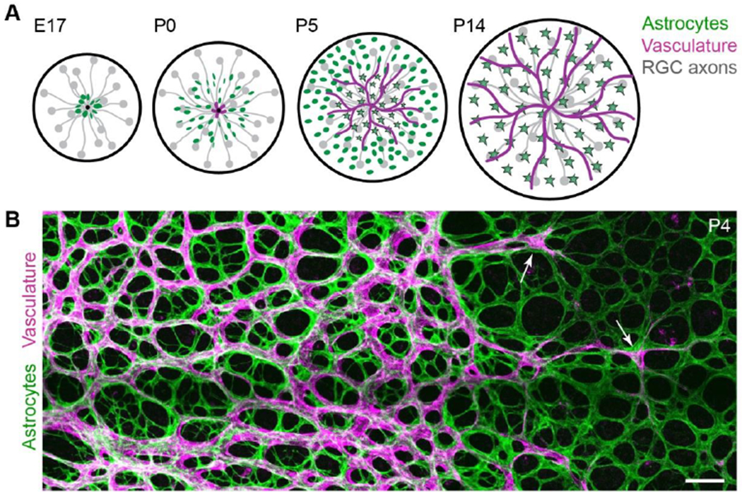
Figure 1: Development of the retinal nerve fiber layer.

A) Schematic detailing sequential development of cell types comprising the RNFL. Before birth (E17 in mice), astrocytes begin to migrate into the RNFL using RGC axons as guides. Vessels enter postnatally (P0). Astrocytes mature as vessels pass over (stars, mature astrocytes).
B) En face confocal photomicrograph of flat-mounted retina depicting RNFL angiogenesis. Incoming vasculature (magenta; labeled by Griffonia simplicifolia Isolectin B4) progresses towards peripheral retina (at right), directly following the preformed astrocyte network (green, anti-PDGFRα). Arrows indicate tip cells at the angiogenic vanguard. Scale bar, 100 μm. (A) modified from Puñal et al. (2019); (B) modified from Perelli et al. (2021).
