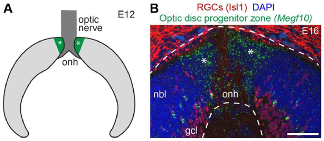
Figure 3: Retinal astrocytes are produced by specialized neuroepithelial progenitors.

A) Schematic depicting cross-section of embryonic mouse retina. The optic disc progenitor zone (green, asterisks), which gives rise to retinal astrocytes, surrounds the optic nerve head (dark gray). Light gray, retinal progenitors derived from the optic cup; these give rise to retinal neurons and Müller glia.
B) Photomicrograph of E16 mouse retinal cross section. In situ hybridization for Megf10 gene, an astrocyte marker, reveals location of optic disc progenitor zone (asterisks). Dashed lines demarcate neural retina. Anti-Isl1 staining shows location of RGC cell bodies (also note non-specific staining outside neural retina).
Abbreviations: onh, optic nerve head; nbl, neuroblast layer containing retinal progenitors; gcl, ganglion cell layer. Scale bar, 50 μm.
