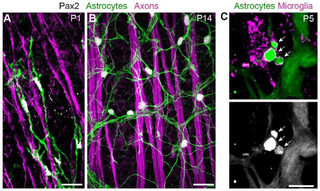
Figure 4: Cell-cell interactions of retinal astrocytes.

A,B) Interactions with axons (labeled by anti-Neurofilament medium chain).
At P1 (A), astrocyte precursor cells climb along RGC axons as they migrate towards peripheral retina. Note polarization of astrocyte arbors (revealed by Pax2-Cre-driven membrane-GFP reporter) along the axon bundles.
At P14 (B), mature astrocytes are no longer closely associated with RGC axons, instead showing uniform “mosaic” spacing. Astrocyte arbors labeled by anti-GFAP. See O’Sullivan et al. (2017) for further details on astrocyte-axon interactions during migration.
C) Interactions with microglia. At P5, astrocyte debris (arrows) can be found within microglia. Astrocytes were labeled with GFAP-Cre-driven TdTomato reporter; microglia were labeled with a combination of antibodies to Iba1 and P2Y12. At this age, GFAP-Cre is selective for astrocytes and not yet expressed by Müller glia.
Scale bars: 25μm (A,B), 10μm (C). (C) modified from Puñal et al. (2019).
