Fig. 1.
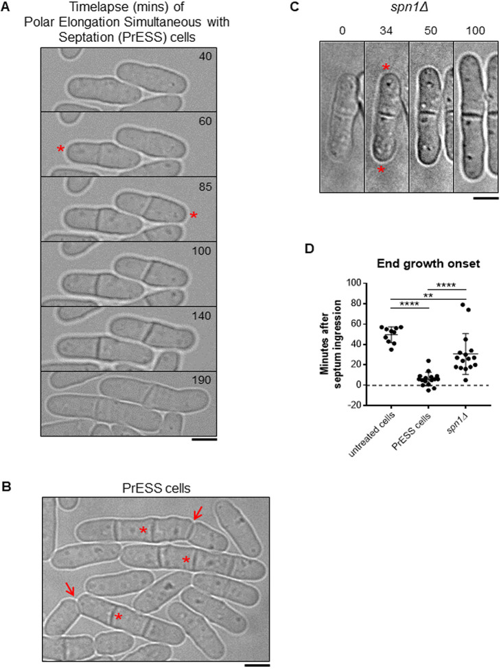
Cytokinetic delay uncouples end-growth resumption from cell separation. (A) Time lapse of cells displaying the PrESS phenotype after LatA washout. Growth initiation is indicated by asterisks. (B) Asynchronous wild-type cells were treated with 10 µM LatA for 30 min, then washed and allowed to recover. A subset of cells show the PrESS phenotype, in which they resume growth as the septum forms and eventually fail to separate (asterisks next to original septum). In the next cell cycle, these cells grow and separate normally (arrows). (C) Time lapse of growth after division in an spn1Δ cell. Asterisks denote growth initiation. (D) PrESS cells resume growth significantly earlier in relation to septum closure than untreated or spn1Δ cells (ordinary one-way ANOVA with Tukey’s multiple comparisons test; **P=0.004, ****P<0.0001; n≥10 cells). Timestamps in B and C refer to time since completion of septum closure. Scale bars: 5 μm.

