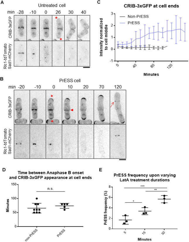
Fig. 3.
Cdc42 is activated at the ends prior to cell separation in PrESS cells. (A,B) Localization of the active Cdc42 marker CRIB-3xGFP in an untreated cell (A) and a PrESS cell (B). Asterisks denote growth onset. Arrowhead denotes loss of CRIB-3xGFP from the cell middle. Arrow shows new ring formation in the next cell cycle. Rlc1-tdTomato and Sad1-mCherry mark the actomyosin ring and spindle pole bodies. Time ‘0’ marks ring/septum closure. A single z-plane is shown for clarity and hence the spindle pole body is not visible in all the time frames. (C) Quantification of CRIB-3xGFP intensities at cell ends of non-PrESS and PrESS cells, normalized to the cell middle. n=6 cells. (D) The time between anaphase B completion and CRIB-3xGFP appearance at the ends is similar in non-PrESS and PrESS cells. (E) The PrESS frequency is lower in cells treated with LatA for a shorter duration than in cells treated for a longer duration. n>800 cells. An ordinary one-way ANOVA with two-stage step-up method of Benjamini, Krieger and Yekutieli multiple comparisons test was used for analysis (n.s., not significant; *P=0.0246; **P=0.0073; ***P=0.0004).

