Fig. 6.
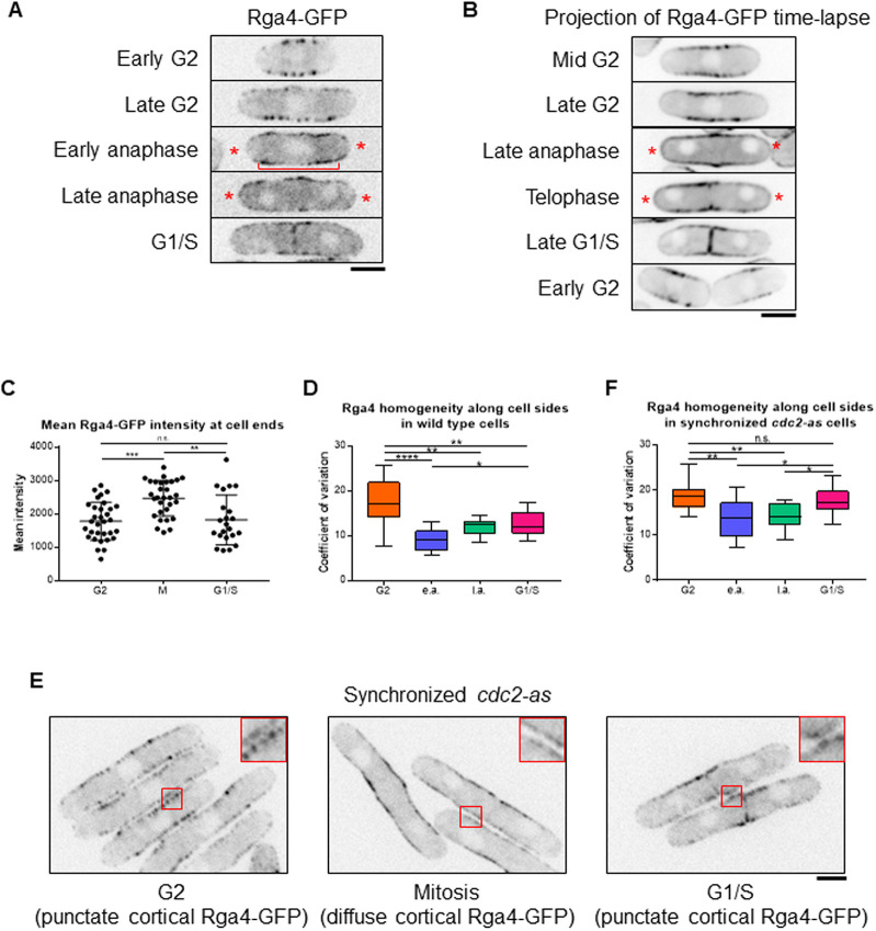
Rga4 localization changes in a cell-cycle-dependent manner. (A) During G2 and septation (G1/S), Rga4-GFP localizes to the cortex along the cell sides as distinct puncta. During late and early anaphase, Rga4-GFP at the cortex appears diffuse (bracket) with localization extending to the ends (asterisks). (B) Time-lapse projection (10 s intervals over 5 min) of Rga4-GFP localization at different cell-cycle stages. Asterisks denote end localization in mitotic cells. (C) Quantification of Rga4-GFP intensities at cell ends during different cell-cycle stages. Significantly more Rga4-GFP localizes to cell ends during mitosis than during either G2 (P=0.0002) or G1/S (P=0.0027). (D) Quantification of Rga4-GFP localization pattern along cell sides (increased co-efficient of variance=decreased homogeneity=increased punctate appearance) in wild-type cells through different cell-cycle stages (n≥10 cells). e.a., early anaphase; l.a., late anaphase. (E) cdc2-as mutant cells blocked in G2 upon 1NM-PP1 treatment show distinct Rga4-GFP puncta along cell sides. After 1NM-PP1 washout, cdc2-as mutant enters mitosis and cortical Rga4-GFP appears more diffuse. In G1/S, Rga4-GFP again localizes as puncta. (F) Quantification of Rga4-GFP homogeneity along the sides, as done for D, in synchronized cdc2-as cells through different cell-cycle stages (n≥14 cells). Ordinary one-way ANOVA with Tukey's multiple comparisons was used for statistical analysis (n.s., not significant; *P<0.05; **P<0.01; ***P<0.001; ****P<0.0001). Scale bars: 5 µm.

