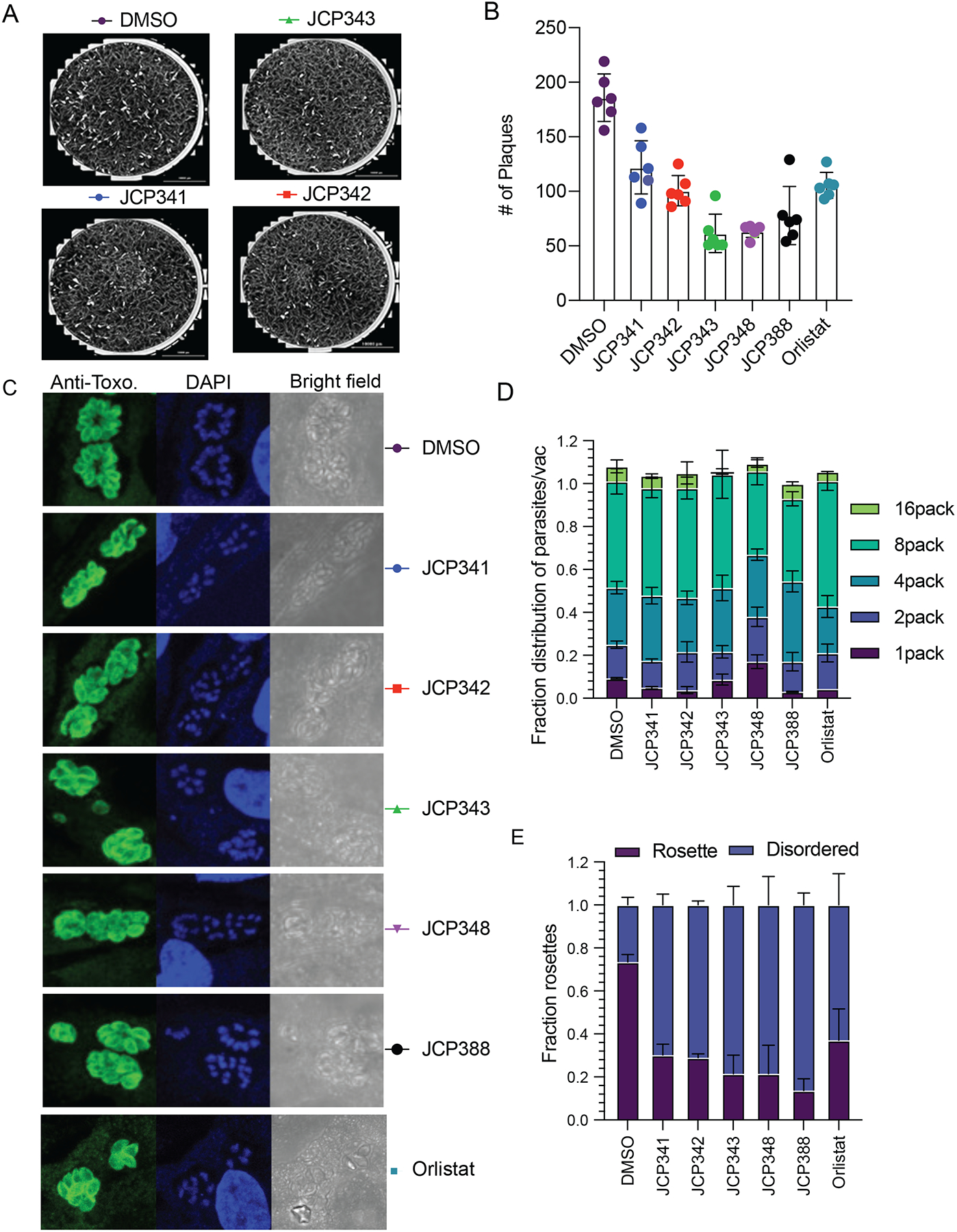
Figure 6. Inhibition of TgASH proteins impairs parasite infection.

(A) Representative images of plaques formed by parasites after pretreatment of the parasites with either DMSO or the indicated screening hits (10 μM final). (B) Quantification of total plaques numbers upon compound treatment. Data represent averages (bar plot) from three independent experiments performed in technical triplicates. (C) Quantification of fractions of parasites per vacuole of parasites treated with either DMSO or selected inhibitors. (D) Indirect immunofluorescence images of wild-type parasites treated with either DMSO or indicated inhibitors. Parasites are stained with an anti-Toxo antibody (green) and nuclei of host cells stained with DAPI (blue). (E) Quantification of fractions of parasites in ordered and disordered rosettes for DMSO or compound treated wild type parasites. The graph presents averaged results from three independent experiments performed in technical triplicate. For replication assays and rosette assay, at least 100 vacuoles/per condition were counted (see also Figure S5).
