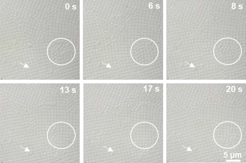
Figure 6.
Brightfield microscopic images of packed ULC microgels assembled into square lattices. As particles diffuse into the defects and grain boundaries, they appear to change from a cubic to a spherical shape, as indicated by the arrows. This shape change can also be clearly seen in the area marked by circle. The scale bar is same for all images.

