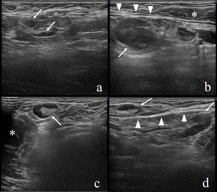
Fig. 1.
A woman in her 30 s with no medical antecedents. a The normal axillary lymph nodes on screening ultrasonography (arrows). b Nine days later, targeted sonography revealed a left axillary lymph node measuring 20 × 15 mm (arrow) deeper than the lower edge of the pectoralis major muscle. c Lymph nodes up to 12 × 7 mm were found on the front side of the lower edge of the pectoralis major muscle (arrow). d In a follow-up ultrasonography 14 days after vaccination, the lymph nodes shrank slightly (arrows). Arrowheads, the hyperechoic line of the lower edge of the pectoralis major muscle; *the pectoralis major muscle

