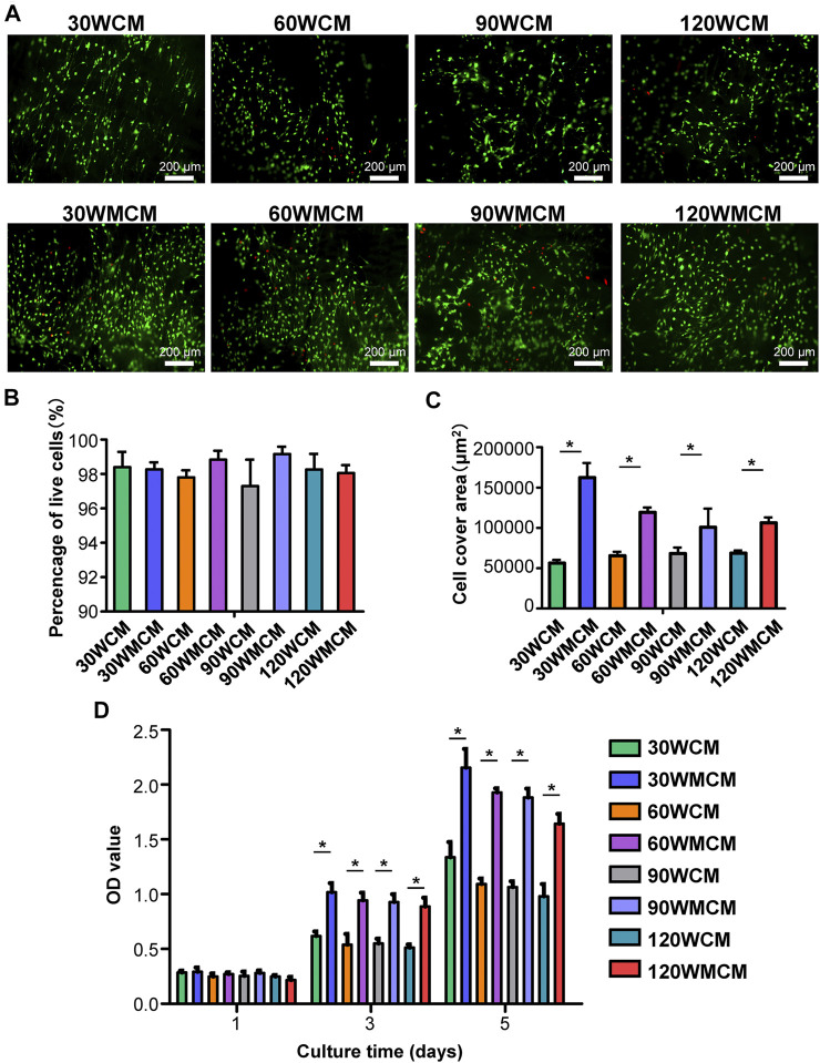
FIGURE 6.
(A) Representative live/dead images of cells cultured for 5 days on the unmineralized and mineralized collagen membranes. The live cells were stained green and dead cells were stained red. (B) The percentage of live cells and (C) cell cover area calculated from live/dead images of cells. (D) The CCK-8 results of cell proliferation on days 1, 3, and 5. The asterisk on top of the bar indicates a statistically significant difference between groups (p < 0.05).

