Figure 4.

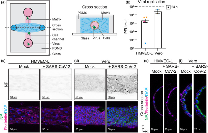
Endothelial cells are not infected with 2 × 10⁶ PFU of SARS‐CoV‐2 in 3D flow‐pressured tubes. (a) Schematic of microfabricated device used to culture 3D vessel tubes under oscillatory flow. The cross‐section shows the lumenised tube covered with endothelial cells surrounded by extracellular matrix. Virus can be flowed through the lumen of the tubes. (b) Viral replication shown as number of PFU mL−1 of supernatant from SARS‐CoV‐2‐infected HMVEC‐L and Vero cells at 24 h after infection. n = 3 (HMVEC‐L), n = 2 (Vero) independent experiments. Representative immunofluorescence images of (c) HMVEC‐L and (d) Vero stained for nucleocapsid protein (NP) (shown as single channel in top panel) (green), Phalloidin (magenta) and DAPI (blue) with mock or SARS‐CoV‐2 infection in the lumen of the tube at 24 h after infection. Scale bar 50 µm. YZ projection of HMVEC‐L (merged image in c) and (f) Vero (merged image in d), with luminal side towards the right of image and basolateral side towards the left of images. Scale bar 20 µm.
