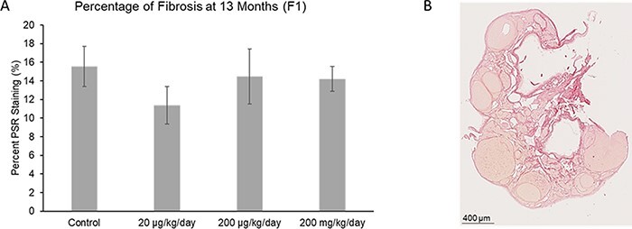
Figure 6:

Effect of prenatal exposure to the phthalate mixture on the percentage of fibrosis in the ovary at 13 months of age in the F1 generation of female mice. Ovaries were sectioned and stained with PSR to evaluate the amount of fibrosis. The percentage of fibrosis was analyzed for each treatment group (panel A: control = 10 females/treatment group, 20 µg/kg/day = 6 females/treatment group, 200 µg/kg/day = 9 females/treatment group, 200 mg/kg/day = 9 females/treatment group). Graphs represent mean percentages ± SEM in the F1 generation of female mice. The image (panel B) shows an ovarian section, with dark red staining indicating fibrosis from a 13 month old mouse in the F1 generation
