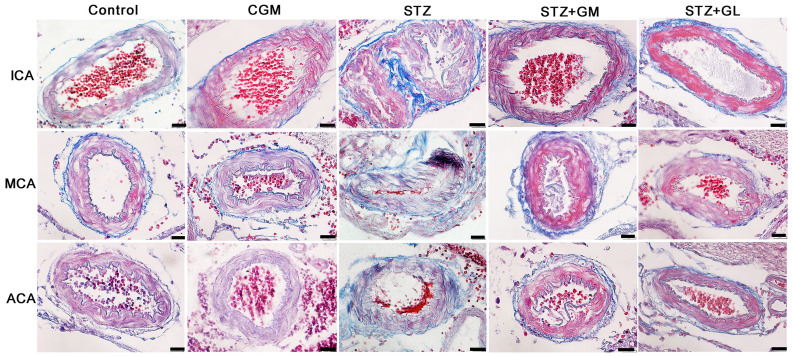
Figure 2.
Photomicrographs demonstrating the histological structure of the anterior arteries included in the circle of Willis. Samples obtained from rats in the C, CGM, STZ, STZ + GM and STZ + GL groups were subjected to Masson's trichrome staining (magnification, x600). ACA, anterior cerebral artery; C, control; CGM, gymnemic control; GL, glibenclamide; GM, gymnemic acid; ICA, internal carotid artery; MCA, middle cerebral artery; STZ, streptozotocin.

