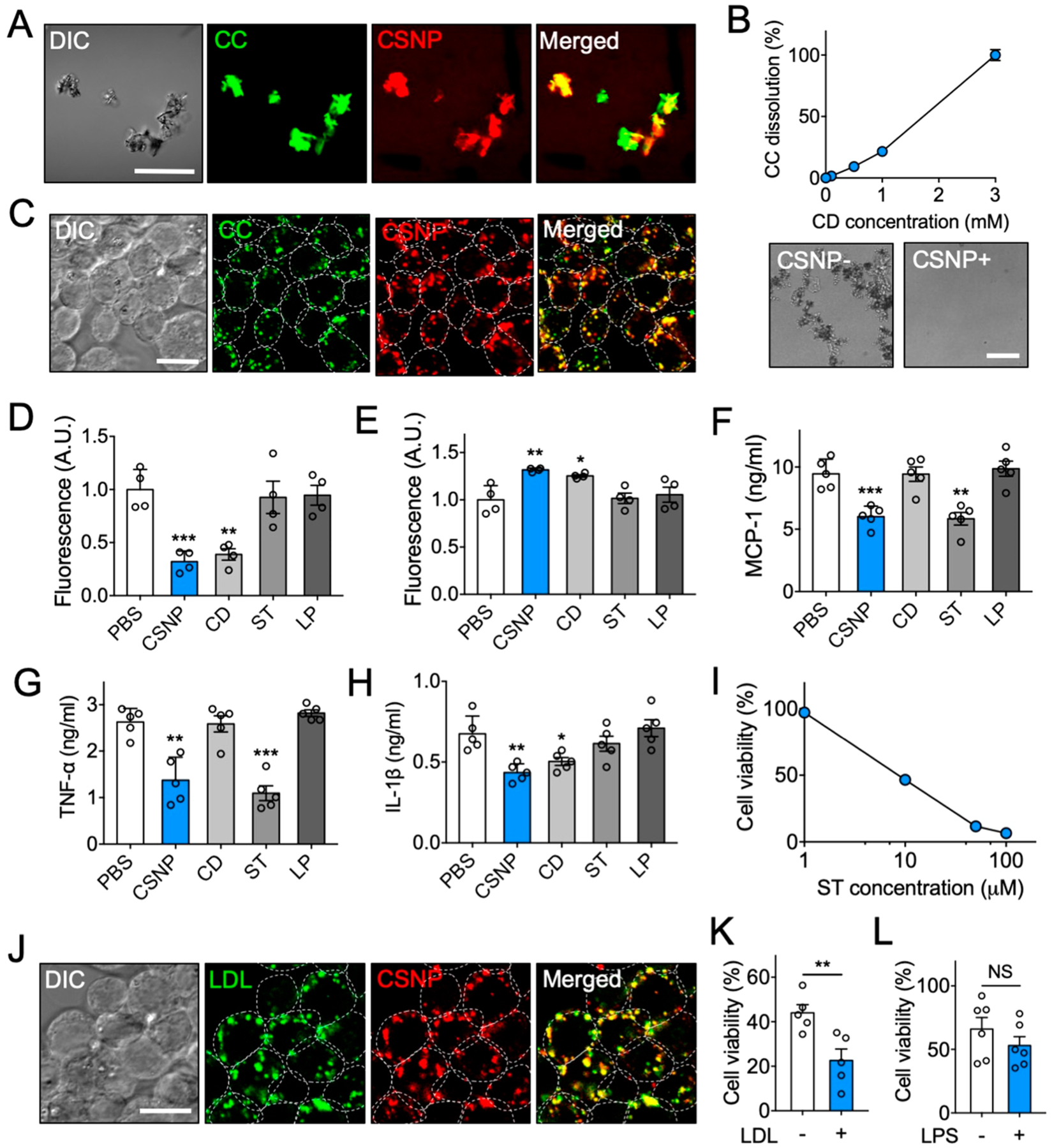
Figure 2.

Multiple anti-inflammatory properties of CSNP in vitro. (A) Representative confocal microscopic images of CSNP binding to cholesterol crystals (CC). The scale bar indicates 50 μm. (B) Dose-dependent CC dissolution with CSNP. Bright-field images show complete dissolution of CC 24 h after CSNP treatment at 3 mM CD. (C) Representative confocal microscopic images of CC-laden macrophages after CSNP treatment. The scale bar indicates 10 μm. (D, E) Assessment of cholesterol (D) within cells and (E) in the supernatant after CSNP treatment. (F, G) Secretion of (F) MCP-1 and (G) TNF-α from the LPS-activated macrophages after CSNP treatment. (H) Secretion of IL-1β from the CC-laden LPS-activated macrophages after CSNP treatment. (I) Dose-dependent effects of CSNP on macrophage viability. (J) Representative confocal microscopic images of LDL-laden macrophage after CSNP treatment. The scale bar indicates 10 μm. (K) LDL-dependent effects of CSNP on macrophage viability. (L) LPS-independent effects of CSNP on macrophage viability. The dotted circles indicate individual cells. Data are mean ± s.e.m. [n = 4 for B–E; n = 5 for F–K; n = 6 for L; NS, not significant, *P < 0.01, **P < 0.005, ***P < 0.001, unpaired two-tailed Student’s t test for K and L, compared with the PBS group for D–H].
