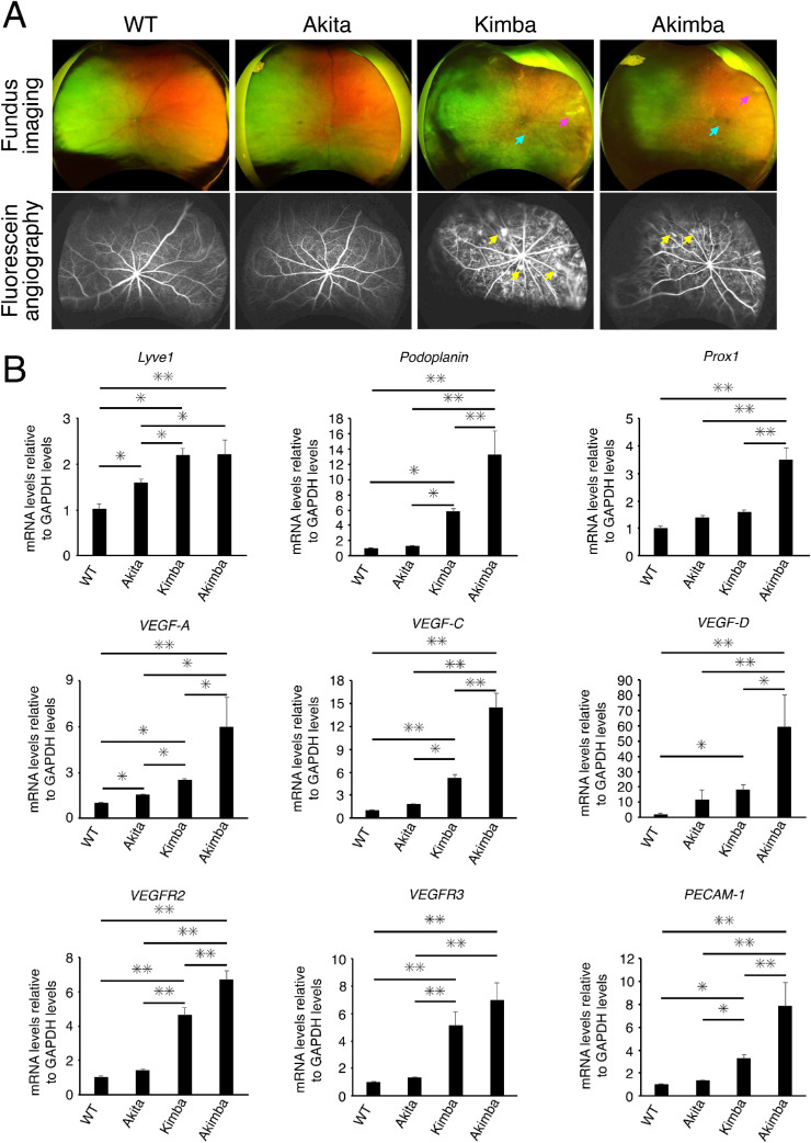
Figure 1.
Characteristics of mouse models of diabetic retinopathy. (A) Representative fundus images and fluorescein angiography (late phase; 2–3 minutes after dye injection) of C57BL/6 WT mice and three different mouse models of diabetic retinopathy (DR) (seven to eight weeks old). These were imaged using an Optos California ultrawide-field imaging system. The three mouse models were heterozygous Akita (Ins2Akita) as a model of type 1 diabetes, and Kimba (vegfa+/+), and Akimba (Ins2Akita vegfa+/−) mice. Blue, pink, and yellow arrows indicate retinal hemorrhage, exudates, and retinal angiogenesis, respectively. (B) Expressions of the mRNAs of various cytokines related to lymphogenesis and angiogenesis in WT, Akita, Kimba, and Kimba retinas (n = 20). Values are the means ± SD. *P < 0.05, **P < 0.01, Student's t-test.

