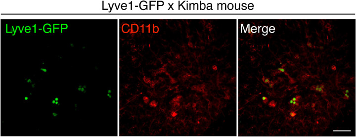
Figure 3.
Population of Lyve1-positive Cells in Kimba Mice. Representative images of flat-mounted retinas of Lyve1-GFP (green) × Kimba mice (vegfa+/+) (Lyve1EGFP/Cre vegfa+/−, 7- to 8-week-old) with staining for CD11b (red). The image shows CD11b cells with GFP expressed in the nucleus. Scale bar: 50 µm.

