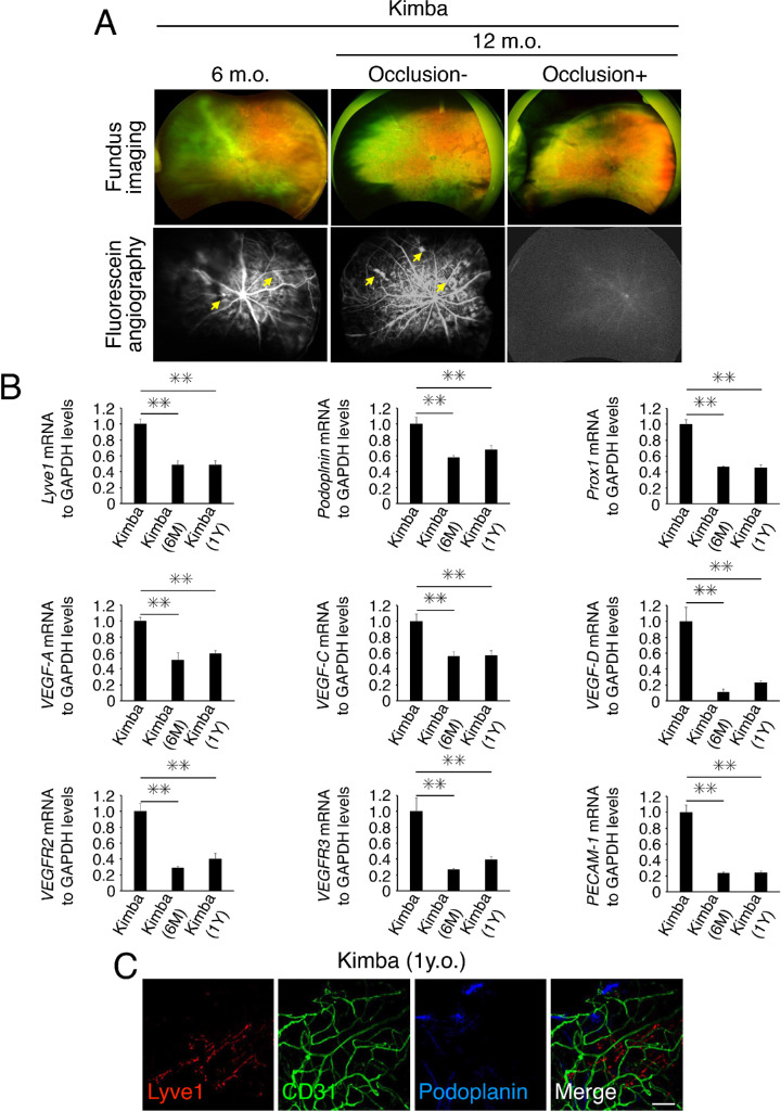
Figure 4.
Characteristics of aged Kimba mice. (A) Representative fundus images and fluorescein angiography (late phase; two to three minutes after dye injection) of C57BL/6 WT mice and three different mouse models of diabetic retinopathy (DR) (6- or 12-month-old). These were imaged using an Optos California ultrawide-field imaging system. Yellow arrows indicate retinal angiogenesis. (B) Expressions of the mRNAs of various cytokines related to lymphogenesis and angiogenesis in retinas from seven- to eight-week-old, six-month-old, and 12-month-old Kimba mice (n = 6). Values are the means ± SD. **P < 0.01, Student's t-test. (C) Representative images of flat-mounted retinas from 12-month-old Kimba mice with staining for Lyve1 (red), CD31 (green), and podoplanin (blue). Scale bar: 50 µm.

