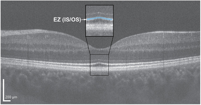
Figure 1.

Horizontal line scan through the foveal center of the left eye of a 24-year-old female with normal vision acquired using a Bioptigen SD-OCT device. Scan was acquired using a setting of 1000 A-scans/B-scan, and the image is a registered average of 20 B-scans. OCT line scans enable delineation of the various retinal layers, including the four hyperreflective outer retinal bands. Of particular interest is the second hyperreflective band, also known as the ellipsoid zone (EZ) or inner segment/outer segment (IS/OS) junction (highlighted blue in the inset black box). Scale bar = 200 µm.
