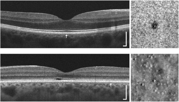
Figure 5.

Comparison of EZ structure on OCT (left) with photoreceptor structure on AOSLO images (right). Top: This 31-year-old female was involved in a motor vehicle collision while traveling on her bike, which resulted in a head injury. The subject's chief complaint was unequal vision, difficulty maintaining clear vision, and photophobia. Clinical OCT imaging (HD-line scan acquired on the Cirrus) did not reveal any disruptions to the outer retina in the right eye. However, when imaged with confocal AOSLO, a small lamellar defect within the foveal mosaic was observed (similar to previous trauma cases).97 This defect was not observed with clinical OCT, likely due to the limited lateral resolution and sampling frequency of the retina with most clinical protocols. Bottom: This 43-year-old female had a family history of progressive vision loss and complained of decreased visual acuity and abnormal color vision. Genetic testing revealed a mutation in the GUCY2D gene (p.R838H; c.251G > A),95 which has been linked to autosomal dominant cone-rod dystrophy. Clinical OCT imaging (HD-line scan acquired on the Cirrus) revealed a focal EZ disruption within the macula of the left eye, while EZ integrity and reflectivity appeared relatively normal beyond the macular region (though there was thinning of the ONL at this location). When a region of the retina was visualized with nonconfocal split-detection AOSLO at ∼5.5° temporal to fixation, it was observed that cone density was significantly reduced (5500 cones/mm2 compared to 12,000 cones/mm2 for normal retinae131). Location of the AOSLO images for each subject is indicated by the arrow in the OCT scan, and they were acquired using previously described protocols.84,95–97 OCT scale bar = 200 µm; AOSLO region of interests are 150 × 150 µm.
