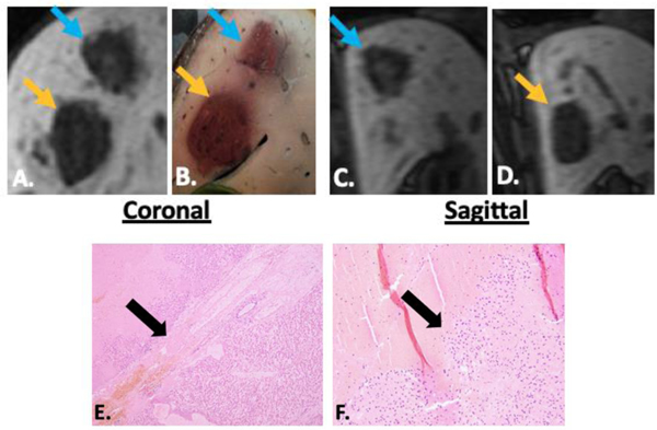
Figure 2.
Delayed MRI imaging in the coronal (A) and sagittal (C, D) planes demonstrates an ellipsoid prescription ablation zone (blue arrow) adjacent to a sphere prescription ablation zone (orange arrow). Correlating gross pathology is seen in B. The narrow transition zone (black arrow) at the border between completely ablated tissue and intact liver is seen in E. (4x) and F. (10x). In the ellipsoid prescription specimens, there was no difference in the diameter of the transition zone from the superior and inferior borders vs. the lateral borders, confirming that the ellipsoid prescription does not extend the zone of partially treated tissue at the cranial or caudal margins.

