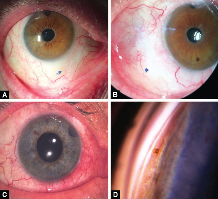
Figs 2A to D.

A) The XEN gel stent (arrow) is visible under the conjunctiva with a flat bleb in the right eye; (B) The appearance of an inferonasally located diffuse filtering bleb (*) in the left eye at month 12; (C) Anterior segment photography after XEN stent surgery in the patient who had undergone two failed trabeculectomies and Ex-Press shunt surgery; (D) View of the XEN gel stent in the iridocorneal angle on gonioscopy in the same patient
