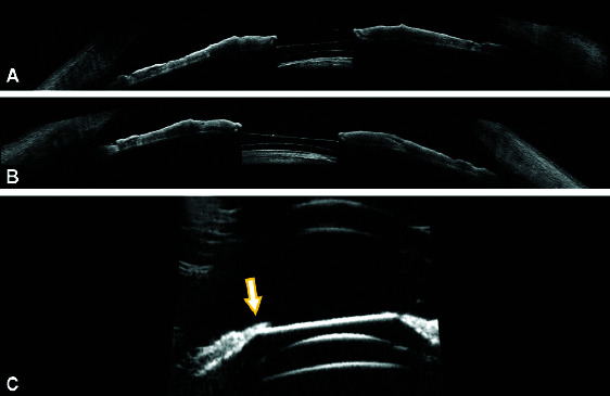
Figs 4A to C.

(A and B) AS-OCT of the right eye and the left eye shows an absence of excess ICL vaulting, shallow anterior chamber; (C) Ultrasound biomicroscopy (UBM) demonstrates direct contact (white arrow) between ICL and the posterior iris in the left eye with the shallow anterior chamber and anterior vaulting of ICL consistent with pupillary block following ICL implantation
