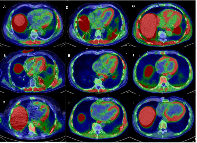
Figure 1.
Fused 68Ga -DOTATOC PET/CT images in the axial plane centered on the long axis of the LV (A–I): patients 3–11 with pathological uptake in the myocardium; pathological uptake in the paravertebral and intercostal muscle is also seen in some patients (A, B, D, E, G); SUV scale 0–2 g/mL. 68Ga -DOTATOC, 68Ga-DOTA(0)-Phe(1)-Tyr(3)-octreotide; LV, left ventricle.

