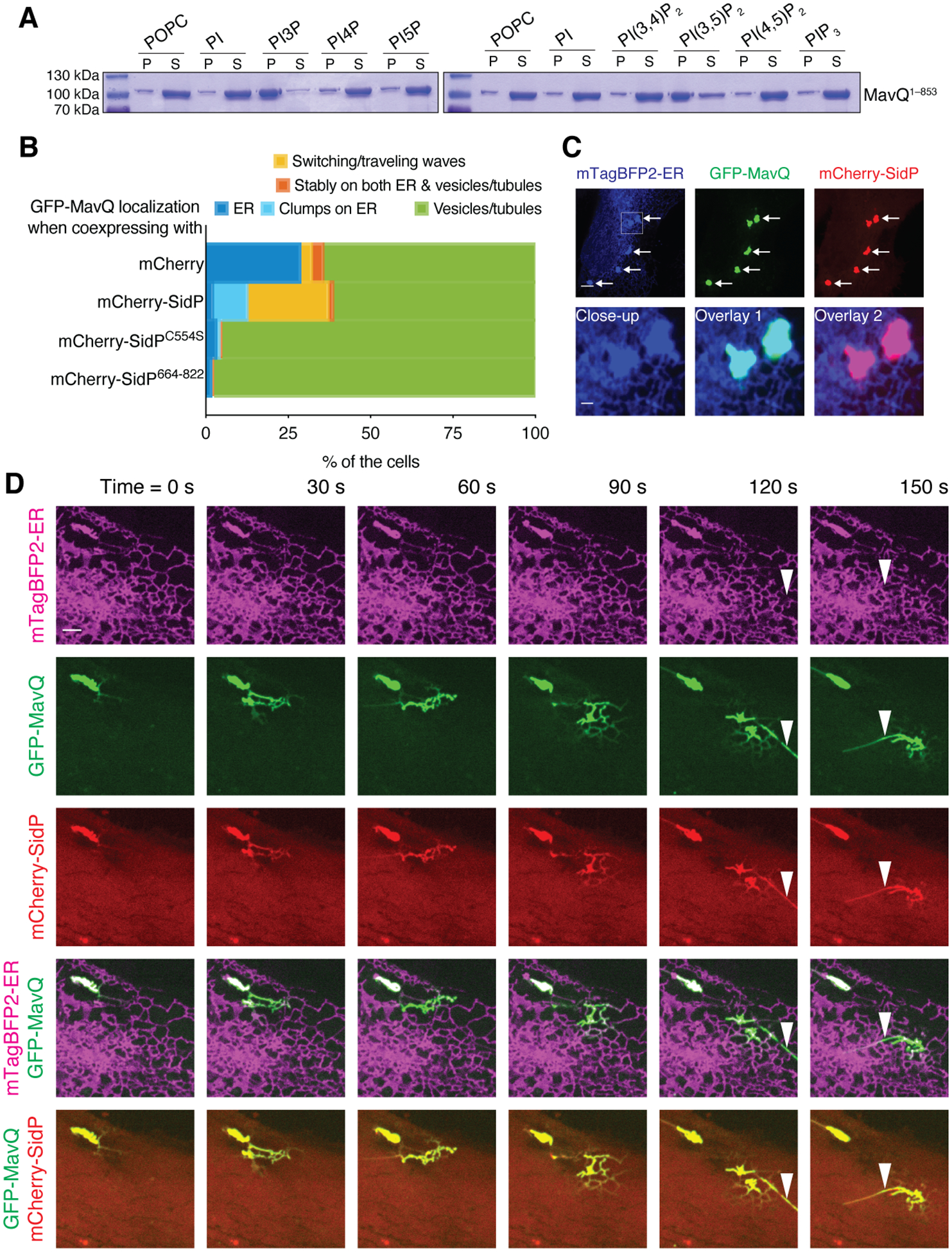
Fig. 4. Positive and negative feedback loops facilitate MavQ protein dynamics and MavQ-induced membrane dynamics.

(A) Sedimentation of MavQ1–853 with POPC or phosphoinositide-containing liposomes. Pellet (P) and supernatant (S) fractions were resolved by SDS-PAGE and visualized by Coomassie staining. Liposome-bound proteins are present in the pellet. See fig. S4C for quantification.
(B) Frequency bar plot showing the proportions of cells with different types of MavQ localization when coexpressed with mCherry, mCherry-SidP, mCherry-SidPC554S or mCherry-SidP664–822 (200 cells/condition pooled from 4 independent experiments). See also fig. S5, B and C.
(C and D) Localization of GFP-MavQ and mCherry-SidP relative to the ER in live HeLa cells by confocal imaging. (C) mCherry-SidP sequesters GFP-MavQ from the main ER network into clumps that still retain ER identity, as indicated by arrows. The close-up shows two such clumps. Bars: 5 and 1 (close-up) μm. (D) Initially, GFP-MavQ and mCherry-SidP concentrate on an ER clump. A wave of ER-bound GFP-MavQ and mCherry-SidP then emits and propagates on the ER network. MavQ/SidP-positive tubules, which are negative for the ER luminal probe mTagBFP2-ER, form during the process (instances marked by arrowheads). See also movie S6. Bars: 3 μm.
