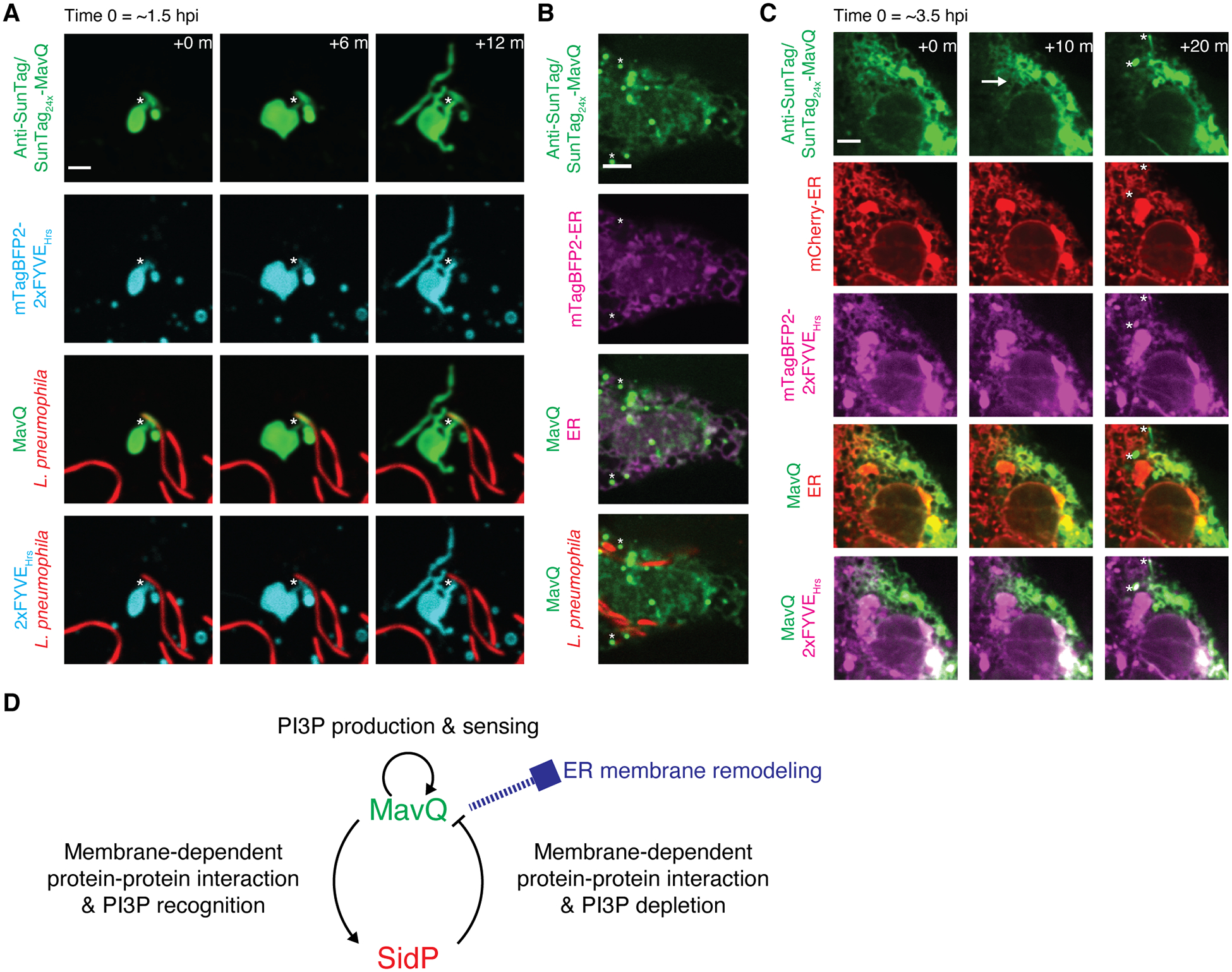
Fig. 5. MavQ exhibits spatiotemporal oscillatory behavior and remodels the host ER membrane during infection.

(A and B) Confocal images of live COS-7 cells expressing anti-SunTag together with the PI3P probe mTagBFP2-2xFYVEHrs (A) or the ER luminal probe mTagBFP2-ER (B) challenged with opsonized, mCherry-labeled L. pneumophila expressing SunTag24x-MavQ. (A) Around 1.5 hr post infection (hpi), MavQ-enriched, PI3P-positive vesicular-tubular structures bud from the LCV. Note that the LCV subdomain positive for MavQ is not PI3P-positive. The neck connecting the LCV and vesicular-tubular structures is marked by asterisks. See also movie S12. (B) Around 5 hpi, MavQ weakly decorates the ER in addition to vesicular-tubular structures (instances marked by asterisks). Bars: 3 (A), 5 μm (B).
(C) Time-lapse confocal images of a live COS-7 cell expressing anti-SunTag, mCherry-ER and mTagBFP2-2xFYVEHrs around 3.5 hpi with opsonized, unlabeled L. pneumophila expressing SunTag24x-MavQ. A wave of MavQ dissociation from the ER occurs (direction of propagation indicated by the arrow), and MavQ-enriched, PI3P-positive vesicles emerge during the process (instances marked by asterisks). Bar: 5 μm. See also movie S15 and movie S16.
(D) Model depicting interlinked positive and negative feedback loops between MavQ and SidP that drive spatiotemporal oscillation of MavQ to facilitate ER membrane remodeling.
