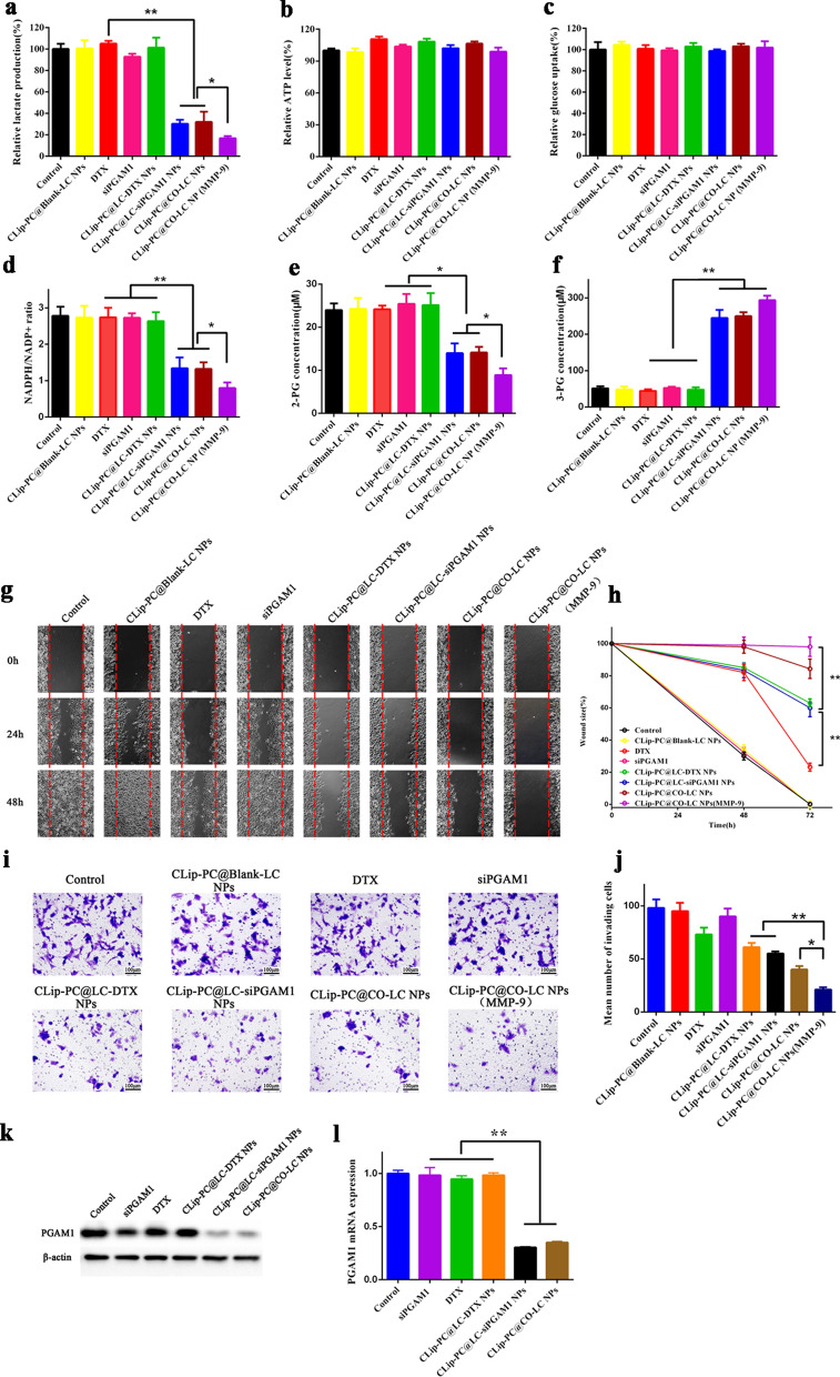
Fig. 4.
Inhibitory effects of CLip-PC@CO-LC NPs on glycolysis, cell migration, and invasion abilities in A549 cells. a Intracellular lactate levels and b ATP levels of A549 cells after 6 h treatment with CLip-PC@Blank-LC NPs, DTX, siPGAM1, CLip-PC@LC-DTX NPs, CLip-PC@LC-siPGAM1 NPs, CLip-PC@CO-LC NPs, or CLip-PC@CO-LC NP (MMP-9). The data are shown as mean ± standard deviation (SD; n = 3). c Normalized glucose uptake of A549 cells after 6 h of incubation with different formulations. The data are shown as mean ± SD (n = 3). d Intracellular NADPH/NADP+, e 2-PG, and f 3-PG were measured after 24 h of incubation with different formulations. The data are shown as mean ± SD (n = 3). g Wound healing analysis of A549 cells treated with CLip-PC@Blank-LC NPs, DTX, siPGAM1, CLip-PC@LC-DTX NPs, CLip-PC@LC-siPGAM1 NPs, CLip-PC@CO-LC NPs, or CLip-PC@CO-LC NPs (MMP-9) for 0, 24, and 48 h; scale bar = 100 μm (mean ± SD, n = 3). h Quantitative analysis of the migration rate in A549 cells for 48 h. i Typical images of Transwell assays of CLip-PC@Blank-LC NPs, DTX, siPGAM1, CLip-PC@LC-DTX NPs, CLip-PC@LC-siPGAM1 NPs, CLip-PC@CO-LC NPs, and CLip-PC@CO-LC NPs (MMP-9) in A549 cells (scale bar = 100 μm). j Quantitative analysis of invading cells in each group. The quantitative data are presented as mean ± SD (n = 3). *P < 0.05, **P < 0.01, ***P < 0.001. k Western blotting assays to determine PGAM1 expression in A549 cells with various treatments. l RT-PCR quantification of PGAM1 mRNA expressed in A549 cells in each group

