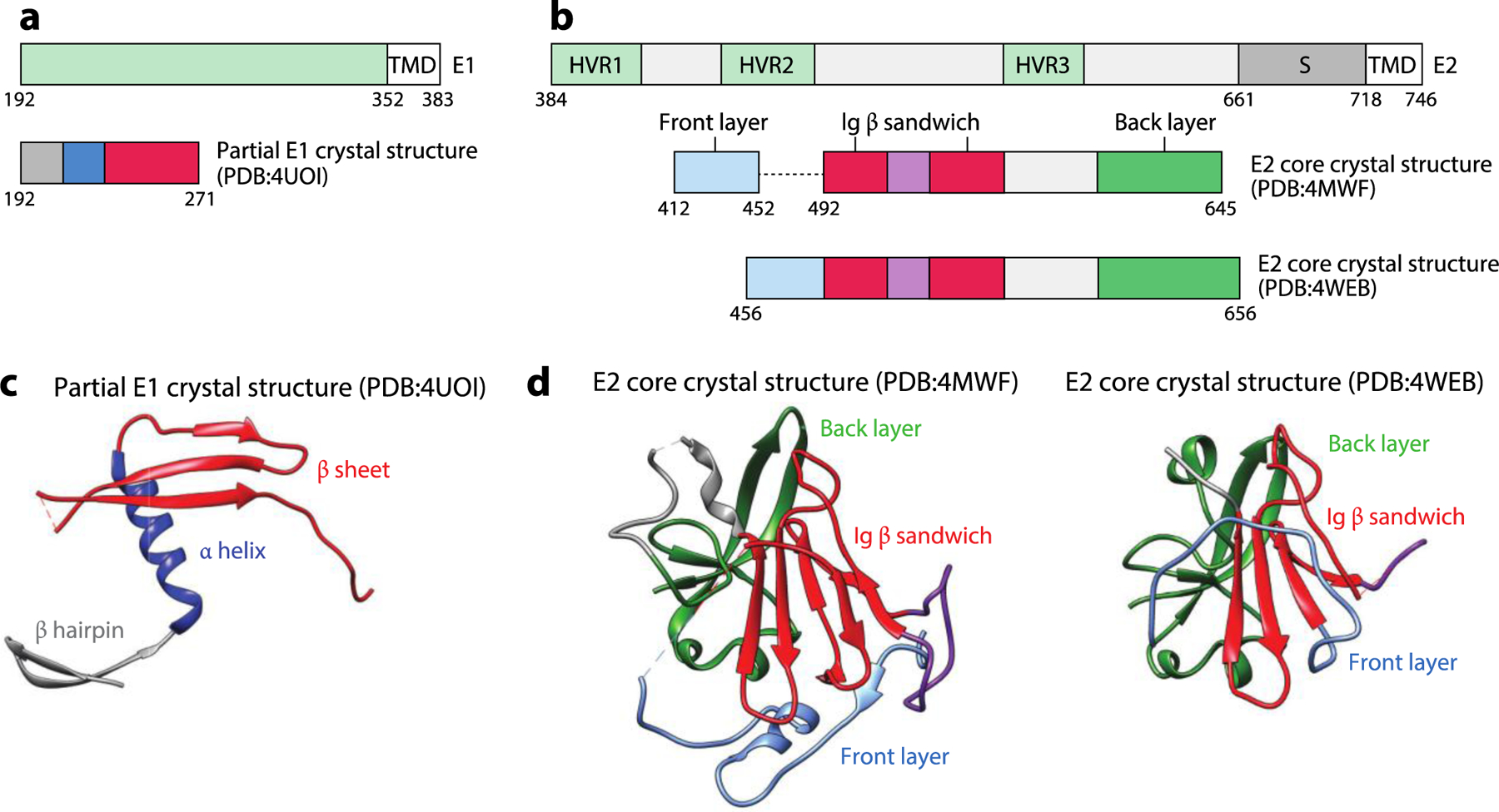
Figure 11.

The HCV E1 and E2 proteins. (a) The schematic diagrams of E1 and the portion of E1 present in the crystallized construct. (b) The schematic diagram of E2 and the portion of E2 present in the crystallized construct. (c) The crystal structure of partial E1 consisting of the N-terminal hairpin, the helix, and a three-stranded β sheet (PDB:4UOI). (d) The crystal structures of the E2 core consist of the front layer, Ig β sandwich, and back layer (PDB:4MWF and PDB:4WEB). Abbreviations: HCV, hepatitis C virus; HVR, hypervariable region; S, stem; TMD, transmembrane domain. Structural graphics were generated with UCSF Chimera, developed by the Resource for Biocomputing, Visualization, and Informatics at the University of California, San Francisco, with support from NIHP41-GM103311 (166).
