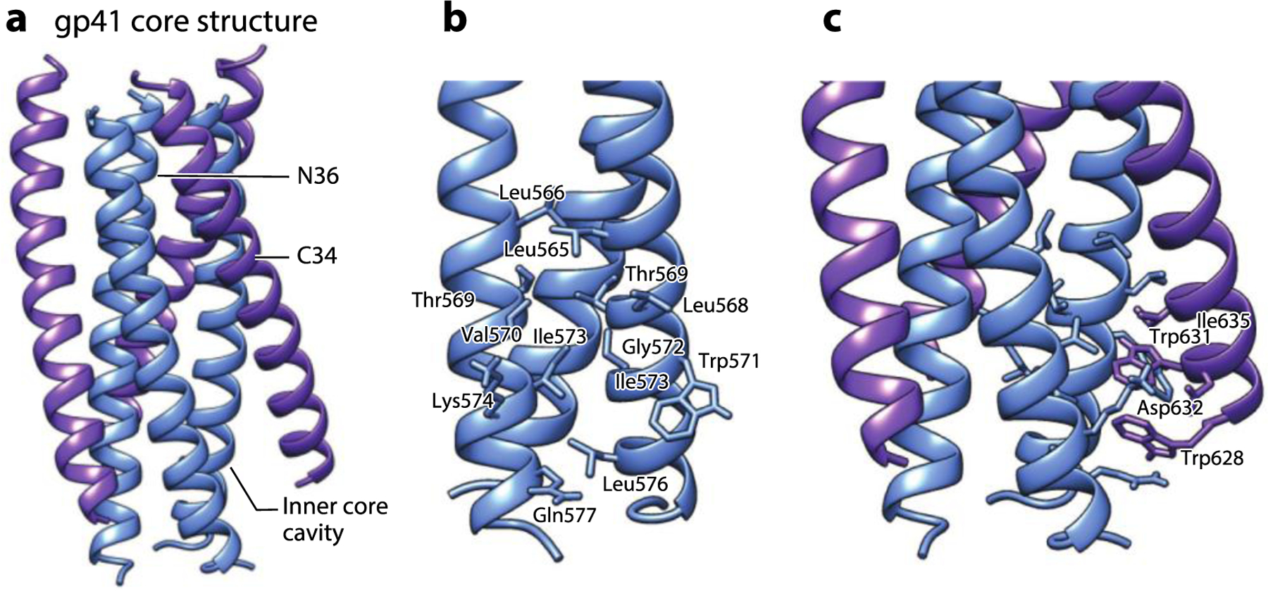
Figure 3.

The gp41 core structure formed by HR1- and HR2-derived synthetic peptides. (a) The six-helix bundle structure formed by N36 (blue) and C34 (purple) peptides derived from HR1 and HR2 of gp41, respectively (PDB:1AIK). (b) The zoomed-in view of the hydrophobic cavity at the HR1 core surface formed by residues from the two adjacent N36 helixes. (c) The zoomed-in view of the residues from C34 interacting with the hydrophobic cavity at the HR1 core surface. Abbreviation: HR, heptad repeat. Structural graphics were generated with UCSF Chimera, developed by the Resource for Biocomputing, Visualization, and Informatics at the University of California, San Francisco, with support from NIH P41-GM103311 (166).
