Figure 2.

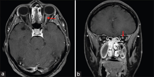
Axial (a) and Coronal (b) T1 weighted contrast and fat saturated MRI sections demonstrate enhancement of the retrobulbar segment of the left optic nerve in a patient with multiple sclerosis

Axial (a) and Coronal (b) T1 weighted contrast and fat saturated MRI sections demonstrate enhancement of the retrobulbar segment of the left optic nerve in a patient with multiple sclerosis