Figure 4.

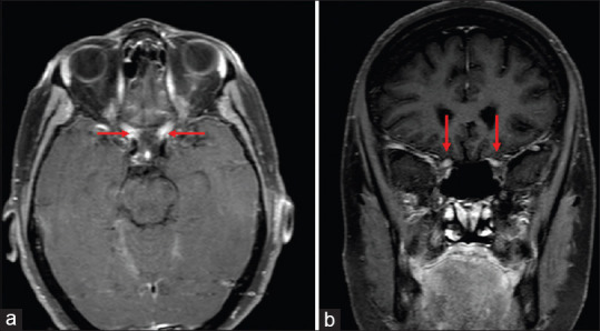
Axial (a) and coronal (b) T1 weighted contrast and fat saturated MRI sections demonstrate edema and enhancement of the both optic nerves from the apex to the optic chiasm in patient with AQP-4 IgG positive NMO disease

Axial (a) and coronal (b) T1 weighted contrast and fat saturated MRI sections demonstrate edema and enhancement of the both optic nerves from the apex to the optic chiasm in patient with AQP-4 IgG positive NMO disease