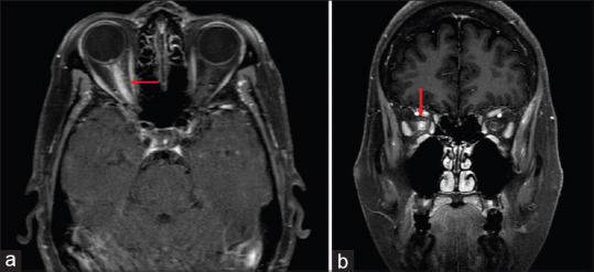
Figure 5.

Axial (a) and Coronal (b) T1 weighted contrast and fat saturated MRI sections demonstrate edema and enhancement of the entire prechiasmatic segment of the right optic nerve. There is also enhancement of the optic nerve sheath and adjacent orbital soft tissue in a patient with MOG IgG optic neuropathy
