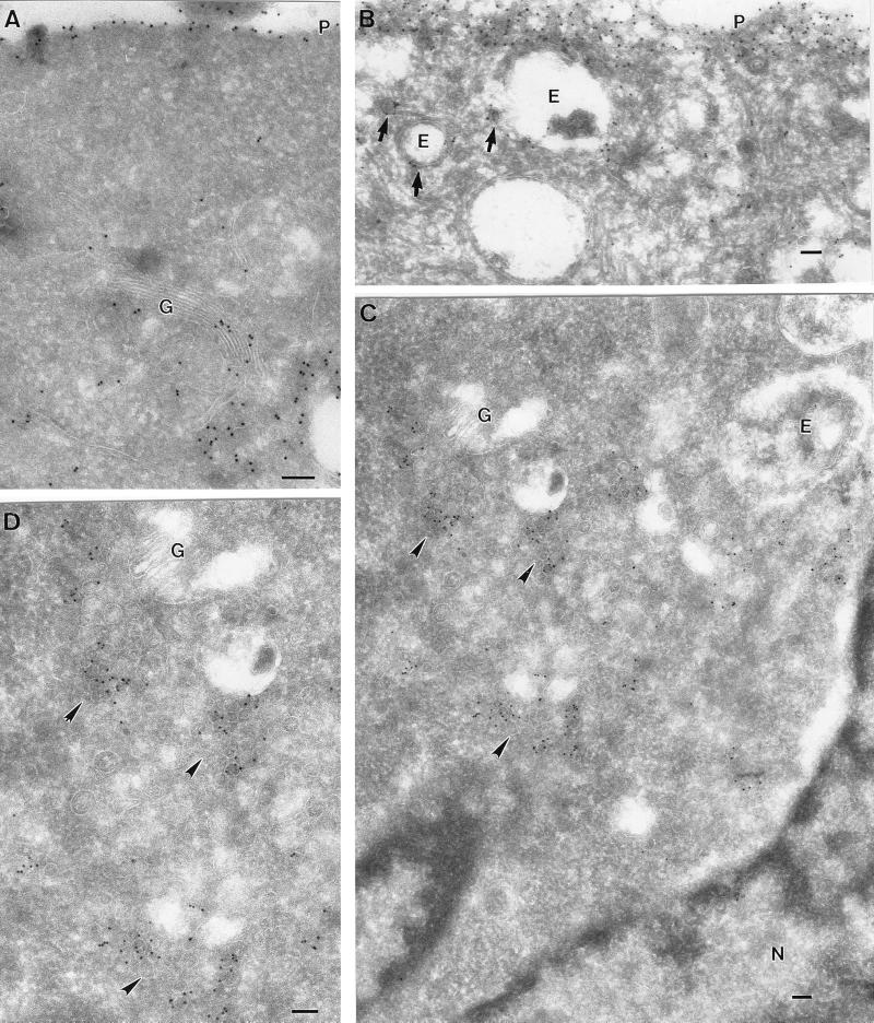
FIG. 3.
Ultrastructural localization of GFP-tH at 37 and 15°C. BHK cells were transfected with GFP-tH and incubated for 18 h at 37°C (A and B) or for 16 h at 37°C, followed by 2 h at 15°C (C and D). The cells were then labeled with antibodies to GFP, followed by 10-nm protein A-gold particles. Scale bars, 100 nm. (A and B) Specific labeling for GFP-tH is evident on the plasma membrane (P). Despite the high expression level, there is negligible cytosolic labeling. Specific labeling is also apparent on the Golgi complex (G). Putative endosomes (E) show low labeling, whereas small vesicles nearby show higher labeling (arrows). (C and D) At 15°C, GFP-tH accumulates in groups of small vesicular structures (arrowheads) close to the Golgi complex (G), as shown at higher magnification in panel D. Again, note the lack of significant cytosolic labeling and the absence of labeling on the ER surrounding the nucleus (N).

