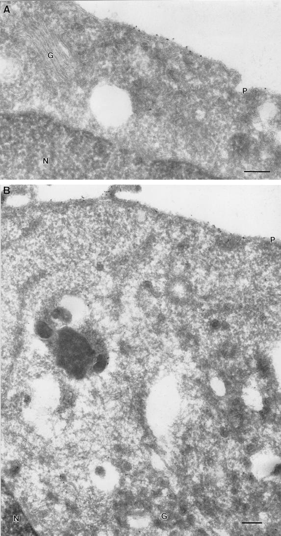
FIG. 4.
Ultrastructural localization of GFP-tK at 37 and 15°C. BHK cells were transfected with GFP-tK and incubated for 18 h at 37°C (A) or for 16 h at 37°C, followed by 2 h at 15°C (B). The cells were then labeled with antibodies to GFP, followed by 10-nm protein A-gold particles. Scale bars, 100 nm. (A) Specific labeling for GFP-tK is evident on the plasma membrane (P). Negligible labeling is apparent on the Golgi complex (G). (B) Again, specific labeling for GFP-tK is evident on the plasma membrane (P). Negligible labeling is apparent on the Golgi complex (G), in contrast to the results obtained with GFP-tH (cf. Fig. 3C and D). N, nucleus.

