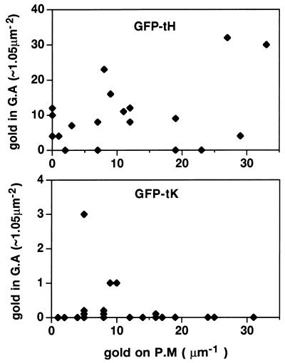
FIG. 6.
Semiquantitative comparison of the subcellular distribution of GFP-ras constructs. Cells expressing GFP-tK and GFP-tH were fixed after a 2-h incubation at 15°C, cryosectioned, and examined by electron microscopy after immunogold labeling for GFP. Cells positively expressing GFPtH or GFPtK were assayed for the number of gold particles present on randomly chosen fixed-unit lengths of plasma membrane (P.M) or in fixed-unit Golgi complex areas (G.A). These data are, in part, arrayed as two scatter diagrams; note the log difference in the y axis scales. The plasma membrane pool, which represents the largest subcellular pool of Ras, contains similar amounts of labeling for GFPtH and GFPtK, indicating that levels of expression were comparable for the two peptides (means, 13.75 and 13.21 gold particles/μm, respectively). GFP-tH was present on and in the vicinity of the Golgi apparatus (mean, 28.62 gold particles/μm2), whereas GFPtK was almost completely excluded from this region (mean, 0.25 gold particles/μm2).

