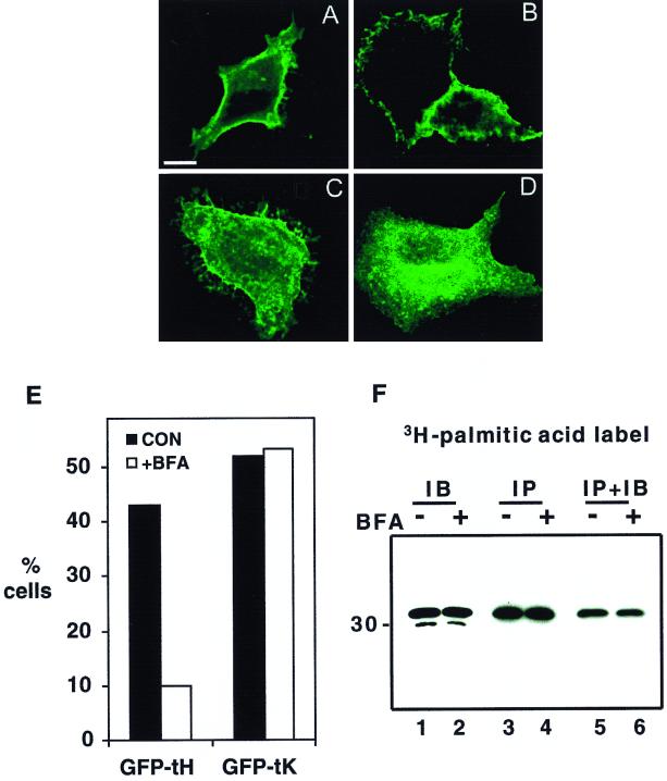
FIG. 7.
BFA inhibits the plasma membrane accumulation of GFP-tH but not GFP-tK. BHK cells were transfected with GFP-tH or GFP-tK. At 2 h after transfection, BFA (5 μg/ml) was added to half of the cultures. After 5 h, coverslips were examined by confocal microscopy. Bar, 10 μm. (A to D) Representative cells from control cultures (A and C) and BFA-treated cultures (B and D). The localization of GFP-tK (A and B) is unaffected, while GFP-tH (C and D) is prevented from trafficking to the plasma membrane. (E) Greater than 450 cells (462 to 524) per experimental condition were scored for intensity of plasma membrane fluorescence by an observer blind to the transfection and treatment conditions. The graph shows the percentages cells with clear plasma membrane staining. It is a representative experiment that was repeated three times with similar results. CON, control. (F) BHK cells plated in 10-cm-diameter dishes were transfected with GFP-tH. At 2 h after transfection, cells were switched to labeling medium containing [3H]palmitic acid; simultaneously, BFA (10 μg/ml) was added to half of the cultures (+BFA) and ethanol carrier was added to the remainder (−BFA). After a further 3 h of incubation, coverslips were quantified as for panel E; the remainder of the cells were harvested into lysis buffer. Lysates were immunoblotted (IB, lanes 1 and 2) or immunoprecipitated (IP, lanes 3 to 6) using monoclonal anti-GFP serum. Radiolabeled proteins in the immunoprecipitates were visualized by fluorography (lanes 3 and 4). Immunoprecipitates were also probed with polyclonal anti-GFP serum to confirm that equal amounts of GFP-tH had been captured (lanes 5 and 6). Note that there is no effect of BFA on GFP-tH expression (lanes 1 and 2) or on palmitic acid incorporation (lanes 3 and 4). BFA did, however, decrease the number of cells with strong plasma membrane staining to 8% from the 48% in the untreated control.

