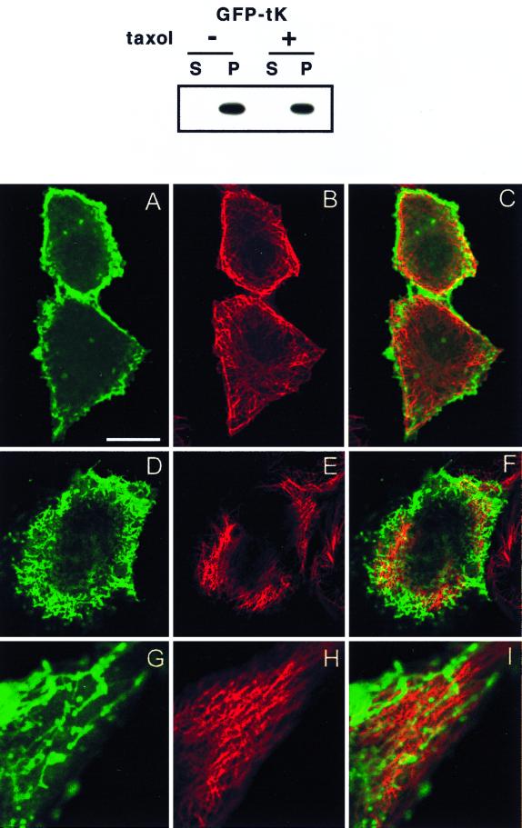
FIG. 8.
Taxol causes redistribution of GFP-tK. BHK cells were incubated in taxol for 2 h prior to and 12 h after lipofection and then harvested for subcellular fractionation or fixed for confocal or electron microscopy. The cytosolic (S = S100) and membrane (P = P100) fractions were prepared from cells expressing GFP-tK as previously described (45), normalized for protein content, and immunoblotted with anti-GFP serum (upper panel). GFP-tK remained fully associated with the P100 fraction in taxol-treated cells. The lower panel shows confocal images of BHK cells from identical cultures that have been costained for α-tubulin. Cells in panels A to F have been cut at the level of the nucleus. The images in panels G to I show the edge of a cell magnified 2.5× relative to the other images. Bar, 10 μm for panels A to F and 4 μm for panels G to I. (A to C) Untreated control cells showing normal microtubules (red channel) and plasma membrane-localized GFP-tK (green channel). (D to I) Taxol-treated cells show some thickening and disruption of the microtubules, accompanied by accumulation of intracellular GFP-tK and a reduction in the amount of GFP-tK seen at the plasma membrane. GFP-tK is seen decorating vesicular and tubulovesicular structures, but there is very little actual colocalization of GFP-tK with tubulin. Some of the tubulovesicular structures are apparently aligned alongside microtubules; this is seen particularly well in panels G to I.

