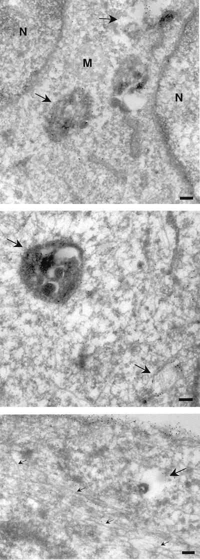
FIG. 9.
Electron microscopic analysis of taxol-treated cells. BHK cells were taxol treated and transfected as described in the legend to Fig. 8. Frozen sections were then labeled with antibodies to GFP, followed by protein A-gold particles. Specific labeling is associated with the plasma membrane and endosomal structures (arrows), but negligible labeling is associated with microtubules (small arrows, bottom panel). N, nucleus; M, mitochondria; bars, 200 nm.

