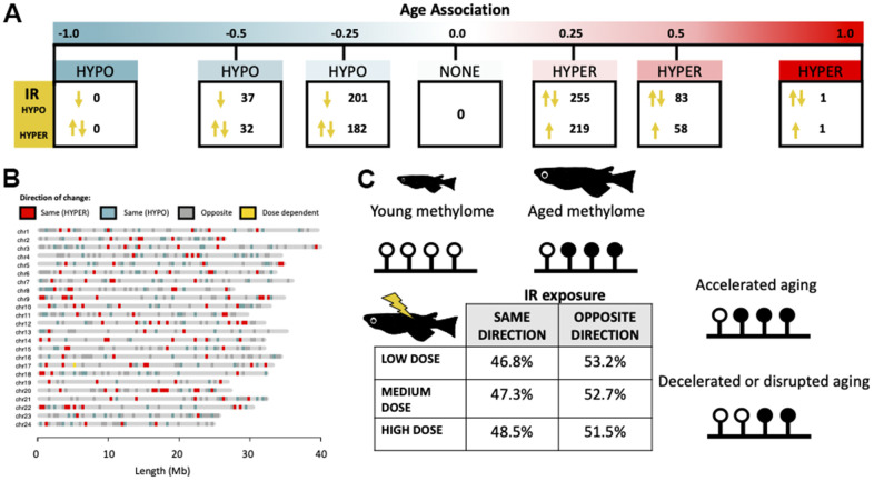
Figure 5.
Directionality of IR-induced changes to methylation status in the context of normal epigenetic aging. (A) Distribution of cytosines which become differentially methylated from IR exposure along the continuum of association with chronological age. Arrows signify whether the IR induced change is in the same or opposite direction as changes induced by age. (B) Genomic distribution of cytosines which become differentially methylated with IR exposure in the same (red/blue) or opposite (gray) direction as age-related changes. Cytosines which become hypermethylated with both age and IR exposure are shown in red and those which become hypomethylated in blue. Cytosines with direction dependent on IR dose are shown in yellow. (C) Table showing the percent of cytosines whose methylation changes in the same and opposite direction across dose rates and a conceptual diagram of the hypothesized effect this could have on the aging methylome.

