Fig. 3.
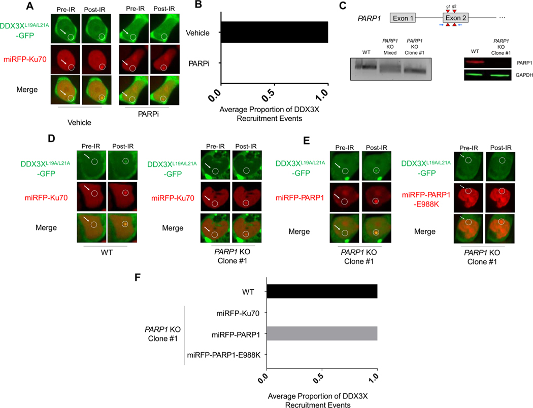
DDX3X recruitment is PARP1-dependent. (A) Representative images of DDX3XL19A/L21A-GFP and miRFP-Ku70 accumulation after microirradiation of HEK293 T cells treated with Olaparib or vehicle. (B) Quantification of accumulation in two independent experiments. (C) Top: Schematic of the PARP1 locus showing the two guide RNA target sites (red triangles) and PCR primers (blue arrows). Bottom: On the right, PCR-based detection of the 70 bp Cas9 deletion at the PARP1 site in the mixed population (middle lane), PARP1-targeted clone (right lane), relative to the parental WT (left lane). Far right, western blot validation of PARP1-deficiency in the PARP1 KO cell line (right lane) compared to the parental WT line (left lane). (D) Representative images of DDX3XL19A/L21A-GFP and miRFP-Ku70 accumulation upon microirradiation (right column) in parental wild-type HEK293 T cells (left) and PARP1 KO cells (right). (E) Representative images of PARP1 rescue with miRFP-PARP1 (red) co-expressed with DDX3XL19A/L21A–GFP (green) upon microirradiation in the PARP1 KO cell line or with the catalytically inactive miRFP670-PARP1-E988 K mutant. The PARP1 rescue experiments were performed three times independently, as quantified in (F).

