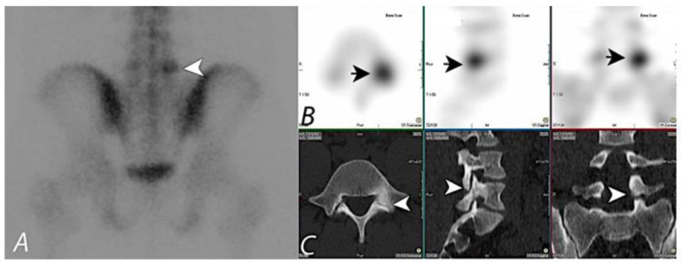
Figure 1.
99mTc MDP bone scan planar (spot) view image (A) of pelvis on a young patient presenting with chronic left sided low back pain, showing focal uptake (black arrowhead) in the lower lumbar region on the left side. SPECT images in axial, sagittal, and coronal planes (B) localizes the activity to posterior aspect of L5 vertebral body on the left side (Short black arrows). Correlative CT images in axial, sagittal, and coronal planes (C) demonstrate a bony defect in the isthmus of the left L5 level pars intraarticularis (white arrowheads), consistent with a monolateral spondylolysis. Taking into consideration the pars defect on anatomic imaging at this level and the focal activity on the bone scan, it can be assumed that the activity most likely correlates with the anatomic abnormality. Lack of co-registration of anatomic and functional information on this set of images prevents definitive interpretation, and hybrid SPECT/CT with fused information can significantly improve diagnostic confidence.

