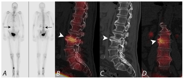
Figure 4.
Anterior and posterior planar images (A) from a 99mTc MDP bone scan on a patient with prostate cancer being evaluated for metastatic disease, with focal uptake along the right aspect of lumbar spine (black arrows). Additional focus of uptake involving the distal right tibia is related to a known fracture involving this region. No additional foci of uptake concerning metastatic disease were identified. Sagittal CT image and fused SPECT/CT images in sagittal and coronal projections (B–D) localize the activity to the right aspect of the disc space between L2 and L3, correlating with disc space narrowing and osteophytosis (white arrowhead). Findings are consistent with uptake related to degenerative disc disease and not related to metastatic disease.

