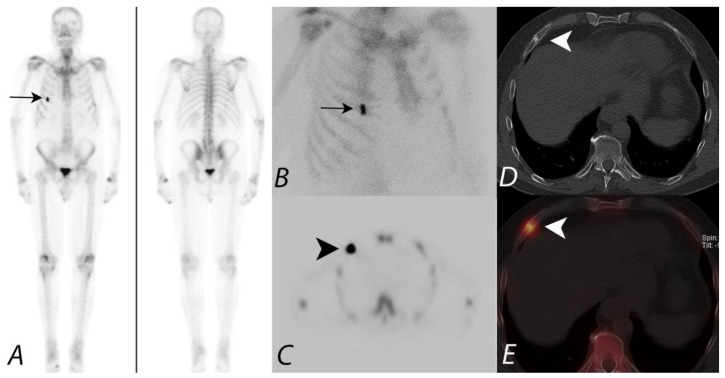
Figure 6.
Patient presenting with prostate cancer underwent 99mTc MDP bone scan to evaluate for metastatic disease. Whole body image in anterior projection (A) showing focal increased tracer uptake along the anterior aspect of the right sided 5th rib, confirmed on planar oblique image (B) (black arrows). SPECT image (C) demonstrates focal uptake along the 5th rib anterior aspect (black arrowhead). However, it is unclear if this focus represents a solitary metastatic disease versus another process, such as trauma. Axial CT (D) at the same level provides anatomic information showing a healing fracture (white arrowhead). Fused SPECT/CT image (E) accurately registers the focal activity (white arrowhead) identified on the planar image and SPECT images to the healing fracture on CT image—a great advantage of the hybrid SPECT/CT imaging combining the functional information with anatomic information.

