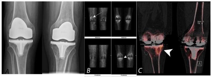
Figure 8.
Bilateral anterior AP radiographs, anterior and posterior planar blood pool and delayed mineral phase images and coronal SPECT/CT images of a Tc-99m MDP bone scan in a 65-year-old male with bilateral painful total knee protheses. Plain radiographs (A) are non-specific (and unchanged over several years) without defined peri-prosthetic lucency. Blood pool images and delayed images (B,C) demonstrate some hyperemia in the joint capsule and serpiginous areas of proximal tibial uptake (white arrows). Clarity is achieved by the SPECT/CT (coronal images, (C)), which demonstrates uptake associated with large, well-corticated areas of bone loss consistent with particle disease (white arrowhead). A similar well corticated region of lucency is also present in the medial aspect of the left proximal tibia.

