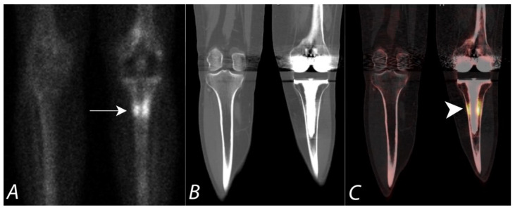
Figure 9.
Fifty-year-old female with a painful left knee prothesis. Anterior planar (A), coronal CT (B) and coronal SPECT/CT (C) images of a Tc-99m MDP bone scan of both knees demonstrate a prominent increased uptake in a band surrounding the midportion of the left tibial prosthetic stem. Plain AP radiographs demonstrate sclerosis without periprosthetic lucency. Findings are consistent with being related to altered biomechanical remodeling (similar to a stress fracture). There is no evidence for loosening or infection.

