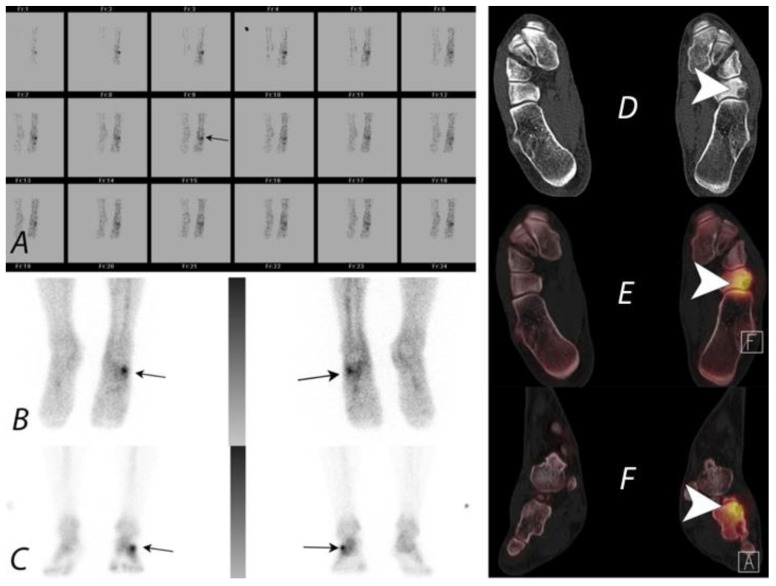
Figure 11.
A young patient presenting with chronic left foot pain underwent three phase bone scan with SPECT/CT. Radiographs were interpreted as normal (not shown). Blood flow and pool images (A,B) demonstrate increased blood flow and pool activity with focal accumulation of activity on the delayed images (C) along the lateral aspect of the mid foot (black arrows). Axial CT image (D) and fused axial (E) and fused coronal (F) SPECT/CT images localize the activity to a well-defined lytic lesion with (white arrowheads) well defined sclerotic rim and a nidus involving the cuboid bone and was interpreted as a possible osteoid osteoma. Patient underwent biopsy, which revealed the lesion to be a benign cartilaginous neoplasm.

