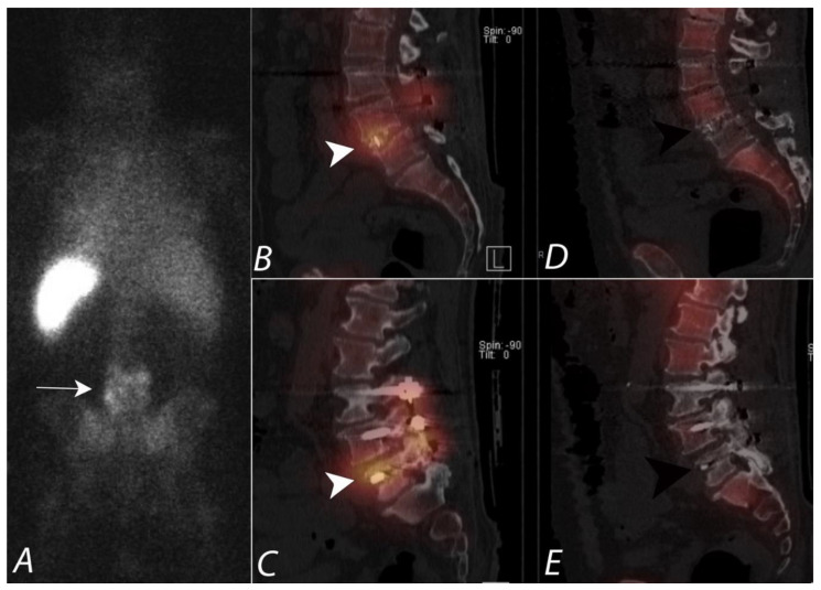
Figure 13.
Seventy-two-year-old male with draining posterior lower lumbar wound one month after dorsal decompression and bilateral dorsal fixation from L3 to S1. Posterior planar image from an 111In leukocyte (WBC) scan (A) demonstrates heterogeneous uptake of tracer activity localizing to the lower lumbar region (white arrow). Sagittal SPECT/CT images of the 111In WBC scan (B,C) show increased uptake of labeled WBCs in the soft tissues, posterior elements, disc space, and vertebral bodies at L4-5. (white arrowheads). However, sagittal SPECT/CT images from 99mTc sulfur colloid scan (D,E) demonstrate no focal uptake (black arrowheads) and is incongruent with the 111In WBC distribution of activity. This supports that a pyogenic infection is present involving the L4-5 vertebrae, posterior elements, and disc spaces. The absence of uptake on sulfur colloid confirms that the uptake of WBCs is not due to a granulation tissue due to mononuclear cell infiltration in chronic inflammation.

