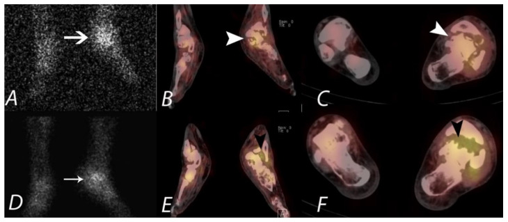
Figure 14.
A seventy-year-old diabetic male patient with chronic left midfoot Charcot joint changes was evaluated for possible superimposed infection. Planar image (A), coronal fused axial image (B), and axial fused axial SPECT/CT image (C) from In-111 WBC scan demonstrate focal uptake localizing to mid and hind foot (large white arrow and white arrowheads), which is congruent with uptake identified on Tc-99m sulfur colloid images (D–F), supporting the presence of chronic inflammation and granulation tissue and not active infection (small arrow and black arrowheads).

