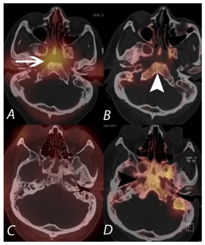
Figure 15.
The upper panel (A,B) demonstrates 67Ga and 99mTc MDP SPECT/CT scans in a patient being evaluated for acute malignant otitis externa. While 99mTc MPD SPECT/CT shows uptake in the bones of the central skull base (white arrowhead), consistent with osteomyelitis, the 67Ga also demonstrates soft tissue infection at the central skull base and around the external auditory canals. The lower panel (C,D) is of a different patient with continued pain following completion of an antibiotic regimen for skull base osteomyelitis. The 99mTc MDP SPECT/CT (right lower panel) shows post-infectious remodeling hyperostosis of the bones of the central skull base, but the 67Ga SPECT/CT (left lower panel) shows an absence of active infection.

