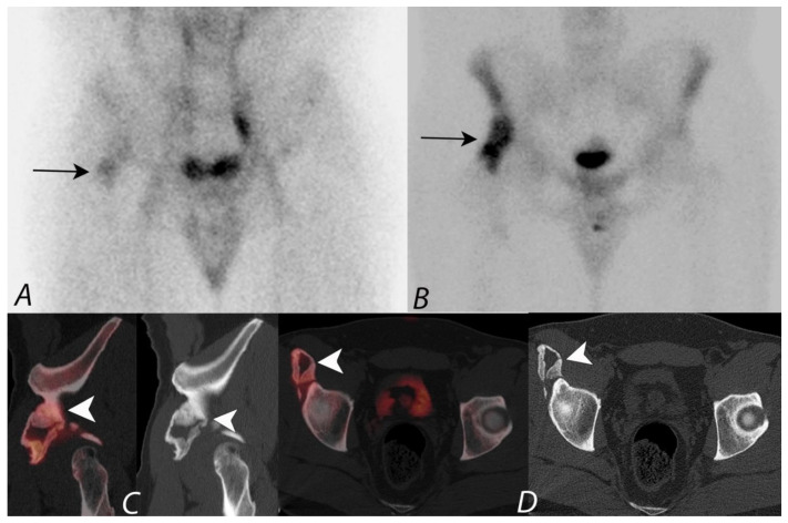
Figure 16.
Patient presenting with right hip and with clinical history of possible heterotopic ossification. Planar blood pool image (A) and delayed image (B) in anterior projection from a 99mTc MDP bone scan demonstrating increased blood pool activity and delayed focal uptake localizing to the right hip region (long black arrows). Fused sagittal image with correlative CT image (C) and fused coronal with correlative CT image (D) localizes the focal activity to an area of exuberant heterotopic ossification along the anterior aspect of the anterior wall of right acetabulum. Given increased blood pool activity, the heterotopic ossification is immature and represents an ongoing process.

