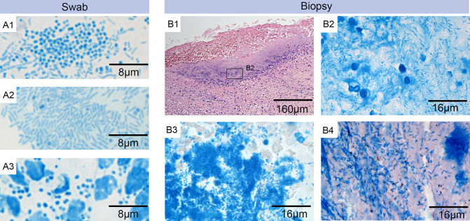
Fig 2. Laboratory analysis.
Microscopic examination of swab smears (A1 –A3) and histological sections (B1 –B4) of biopsies from wounds suspected to be BU lesions. Specimens were either stained with ZN-Methylene blue (A1-A3 and B2-B4) to detect AFBs (pink) and nucleic acid, implicating DNA from secondary infections (blue); or with Haematoxylin-Eosin (B1). A1-A3: Staining of wound exudate from different swabs revealed a heavy infection with rods and/or cocci but no AFBs. A1: rods and cocci (participant 14). A2: rods (participant 11). A3: cocci and leukocytes (participant 4). B1: overview of excised tissue specimen revealing a secondary infection at the open ulcer surface and the underlying tissue (participant 21). B2: higher magnification of this area revealed the presence of rods but no AFBs (participant 21), B3: the same higher magnification at a different area revealed a strong infection with cocci but no AFBs (participant 21) B4: staining of tissue sections from a different participant revealed the presence of large numbers of rods but again no AFBs (participant 6).

